Cancer Treatment X-Ray Tube (ca. 1910-1920)
This unusual looking X-ray tube was designed for use in radiation therapy. Unfortunately, there are no markings on it that might help identify the manufacturer or the date that it was produced.
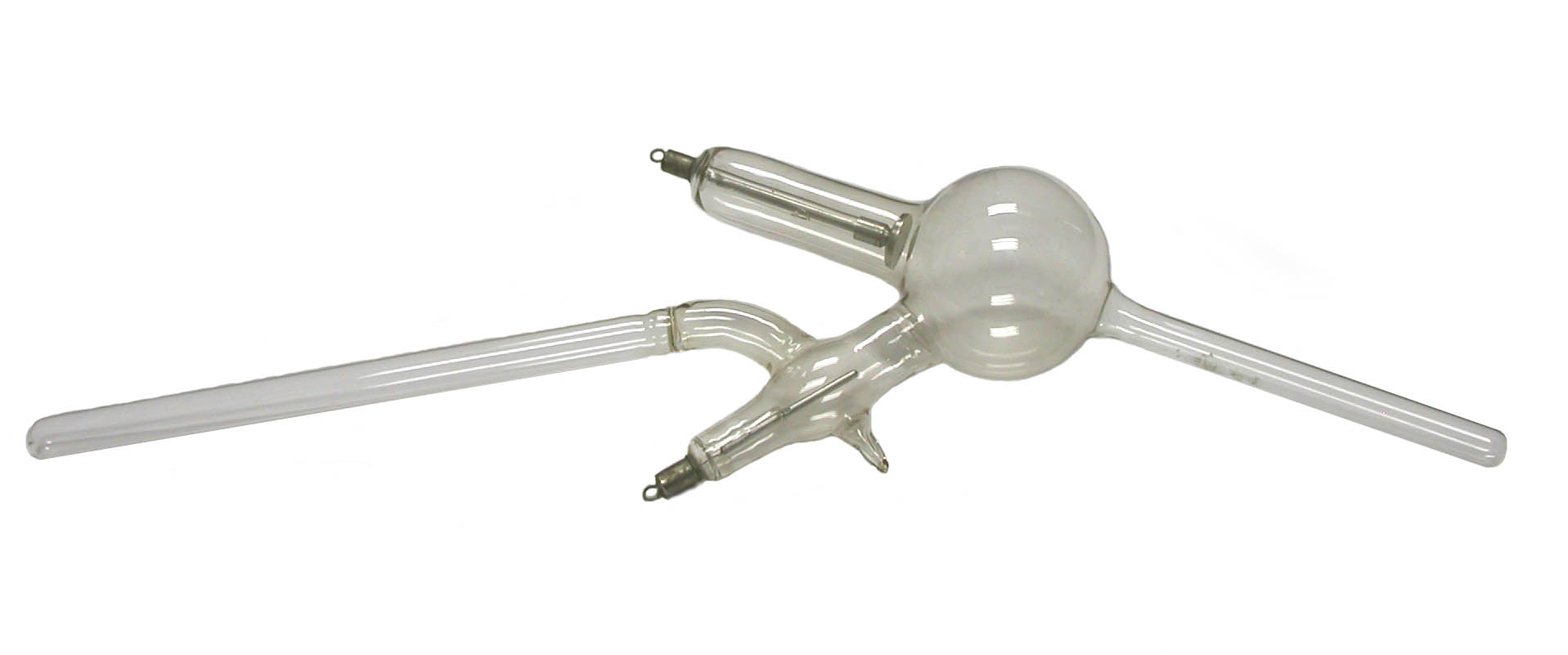
In the photo, a conventional looking cathode can be seen in the glass arm at the point where the latter is attached to the upper left side of the spherical bulb. The other electrode, the anode, is the aluminum rod inside the glass arm attached to the lower left side of the bulb.
Although most X-ray tubes employ metal targets, the target in this tube is glass—more specifically the glass wall of the long narrow extension that projects from the lower right side of the bulb. A somewhat similar extension, attached to the left side of the glass arm enclosing the anode, is the handle!
While the tube was operating, the electrons discharged from the cathode were directed through the large bulb into the extension (pointing down and to the right in the photo) where they generated X-rays as they struck the glass. Compared to the X-rays produced by a typical tube, those produced by this tube were of relatively low intensity and energy. First, it was necessary to employ weak currents so as to prevent the glass (which might be in contact with the patient’s body) from overheating. Second, because the tube was used for superficial rather than deep therapy, low voltages were used in order to keep the energy of the X-rays low.
Tubes of this type were usually made from lead glass except for the extension that served as the source of X-rays. The latter was made from soda glass. When a high voltage was applied to the tube, the two types of glass fluoresced differently: the main body of the tube exhibited a blue color while the extension fluoresced an apple-green color. Of course, the green fluorescence might be difficult to see because the tube was primarily used to treat rectal and vaginal cancers.
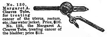
The accompanying undated advertisement refers to this as a “Margaret A. Cleaves” tube. Macalaster-Wiggin also referred to their version of this tube as a "Cleaves' type of cancer tube."
It is worth noting that Margaret Cleaves was the first to employ brachytherapy in the treatment of cancer.
Most references to this type of tube identify it as a “Caldwell” treatment tube, or simply, a cancer treatment tube. An E. B. Meyrowitz catalog (no date) refers to it as the "Caldwell Localizing X-Ray tube."
The following description from a Scheidel-Western X-Ray Coil Company catalog provides some interesting detail:
"This tube is especially adapted for treatment of the vagina, rectum and throat."
"In this tube we have the cathode rays which are thrown along the focus tube, where the X-rays are generated, passing in all directions, both at right angle and parallel to the focus tube."
"The cathode rays are extremely hot, heating the focus part of the tube, and it is necessary to use the water jacket accompanying the tube at all times. The tube is ideal for treating a large or small diseased surface in the vagina or rectum and the X-rays are as high as in a medium vacuum in the regular X-ray tube."
"The time of exposure is reduced and it is unnecessary to shield the patient. The tube is provided with a handle. The water jacket is held in position by a rubber joint."
Unfortunately, the tube in the ORAU collection does not have the water jacket mentioned in the above quote. It does, however, have a great pedigree: it once belonged to M. J. Gross who had worked with Dr. Coolidge at General Electric in Schenectady N.Y. During the 1930s and 1940s, Gross and Zed Attlee formed the core of Coolidge's research and design team. Gross later became Vice President of the GE X-ray Company.
Size: Approximately 20" long with 3" bulb diameter
Kindly donated by Malvern J. Gross Jr. in memory of his father.
References
- Tousey. Medical Electricity and Rontgen Rays. W. B. Saunders Co. 1921.
- Macalaster-Wiggin Co. X-ray Apparatus. 1907.