Water-cooled X-Ray Tube of Unknown Manufacture (ca. 1916-1920)
I would guess that this example of a water-cooled X-ray tube (the only one in the collection) dates from 1916 to 1920 simply because the metal collar around the cathode is stamped 1916. While there is nothing on the tube that would indicate the manufacturer, it is similar in appearance to the drawing of a Macalaster & Wiggin tube in the 1919 text “Radiography and Radio-therapy” by Robert Knox.
For what it is worth, Professor B. Walter at the National Physics Laboratory in Hamburg was the first to suggest the concept of using water to cool the target of an X-ray tube. However, it was the C.H.F Muller company who first produce one—in 1899.
Back in the day, water-cooled tubes were more common in Europe than North America. But they were never very popular anywhere and today they are fairly rare. They certainly had advantages, e.g., they could be operated continuously for hours with high currents and voltages.
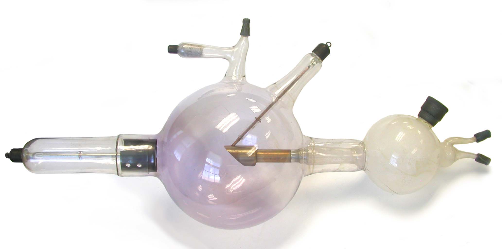
At first glance, I thought that this was a bi-anode tube because of the glass arm extending up out of the bulb at a 45 degree angle. But no, this tube only has two electrodes: the cathode and anode. If it were a bi-anode tube, it would have three: a cathode, an anode and an anticathode. The aforementioned arm projecting at 45 degrees houses the electrical connection to the target. For reasons of safety and simplicity, this was probably better than running the connection through the glass arm connected to the water reservoir. The latter is the smaller glass bulb at the right end of the tube (as seen in the photo).
During use, the reservoir was supposed to be full of water, but some would inevitably evaporate. I believe that it was the evaporation and repeated refilling of this tube's reservoir that was responsible for the buildup of residue on its inside. Even when the tube was in storage, it was common to leave water in the reservoir in order to minimize the chance that it would be dry when the tube was used next. For some unknown reason, operators were recommended to use ordinary tap water rather than distilled or filtered water. Although it is not visible in the photograph, the back side of the water reservoir is severely cracked—that is why it is not visible.
Tubes like this one, with the water reservoir on the long axis of the tube, could be positioned below the couch (i.e., the patient) or above. If the reservoir were projecting out from the tube at a 45 degree angle, a more common arrangement, the tube could only be positioned above the couch. Patients were sometimes nervous about having a glass bulb containing boiling water positioned over their body during a prolonged exposure. Wimps.
The preceding paragraph needs a little clarification. In the early days of roentgenology, it was common to keep the water cool by refilling the reservoir during the exposure. Later, operators tended to follow the suggestion of Bucky (1915) that the water should be allowed to come to a boil. This reduced fluctuations in the tube’s output during the exposure.
The perforated black metal sheath just behind the cathode is very similar to what the Victor X-ray Company called the "Victor supporting protective shield." Its purpose, found in Victor's catalog description of the Snook tube, was to "steady the focus and render the tube practical for use with heavy currents." It was only under heavy loads that enough positive gas ions would strike the cathode for the latter to overheat. As such, this shield might have been designed to remove heat from the cathode and dissipate it through the glass arm. Perhaps more importantly, it would shield the glass closest to the outer edge of the cathode from the heat.
This particular tube belonged to M. J. Gross who worked with Dr. Coolidge at General Electric in Schenectady, N.Y. Gross later became vice president of the GE X-ray Company. During the 1930s and 1940s, Gross and Zed Attlee formed the core of Coolidge's research and design team.
Size: Approximately 7" diameter bulb, 23" long
Kindly donated by Linda Sinrod in memory of her father, Malvern J Gross.
References
- Knox, R. Radiography and Radio-Therapeutics.1919.
- Ronne, P. and Nielsen, A.B.W. Development of the Ion X-ray Tube. 1986.