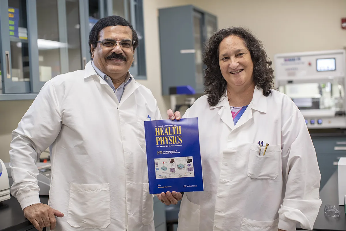
Adayabalam Balajee, Ph.D., and lead biologist Maria B. Escalona hold a copy of the 2023 Health Physics Journal in which their research is published.
How do doctors and scientists assess the health risks of diagnostic low dose radiation exposures such as X-rays? Up until recently, it was a challenging task due to the lack of appropriate model systems that mimic tissue microenvironments. With funding provided by the ORAU-Directed Research and Development program, Adayabalam Balajee, Ph.D., director of ORISE’s Cytogenetic Biodosimetry Laboratory—which is part of DOE-NNSA's Radiation Emergency Assistance Center/Training Site (REAC/TS)—is collaborating with Yong Huang, Ph.D., and his research group at the University of Florida to answer this question. They are developing a three-dimensional human tissue, similar to bio prints, that can be used for the realistic assessment of low-dose radiation effects in a tissue like microenvironment.
“Human exposure to low doses of radiation—especially through medical diagnostics—has become inevitable, and that’s where potential health risks assume a greater importance,” Balajee explained. “Also, sporadic instances of diagnostic overexposures emphasize the need for understanding how low dose radiation exposure affects our tissues and organs and what would be the health consequences in the long run.”
While radiation biology isn’t new—it was discovered in the late 1800s with the use of X-rays—the use of 3D bioprinting technology to evaluate low dose radiation induced effects in a tissue microenvironment is highly innovative and novel.
Also leading edge, tissue engineering emerged around the turn of the millennium and has become more sophisticated in approach and application in recent years. For this particular project, Huang and his research group adapted his design-engineering process toward radiation exposure research.
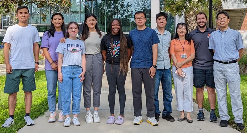
Yong Huang, Ph.D., and his University of Florida research group develop and improve printing technologies for the fabrication of 3D tissue constructs.
Much like 3D printing in an advanced manufacturing environment, developing tissue constructs for research involves combining multiple layers of cellular and other gel matrix material until a three-dimensional human equivalent tissue is created upon which experiments and assessments can be performed. To make a 3D tissue construct, scientists need several millions of cells. In this initial study, the scientists used human foreskin fibroblast (BJ1 human skin cells) immortalized with a telomerase reverse transcriptase (TERT) enzyme that gives them an infinite in vitro life span.
“We aim to develop and improve various printing technologies for the fabrication of 3D heterogeneous soft structures and constructs (with or without cells) made from difficult-to-print materials or fluids using ink jetting, laser-induced forward transfer, extrusion, and/or stereolithography,” Huang said. “Our printed tissue constructs may provide a unique in vitro platform to study some interesting cellular and/or molecular biology problems including cellular response mechanisms to low dose radiation that we are currently investigating.”
Previously, scientists looked at individual cells or a monolayer of 2-dimensional cells to determine radiation-induced damage. However, as Balajee explained, judging the reaction of individual cells to damage is less accurate than studying the damage in a tissue because of the diverse intercellular communication in a tissue context is relevant for predicting potential low-dose, radiation-induced health risks including cancer.
“Intercellular communication is the greatest in a 3D tissue microenvironment relative to 2D monolayer of cells,”Balajee said. “Experimental studies have shown less induction of DNA damage and gene mutations in 3D systems compared to 2D systems.”
It took Huang’s team about six months of trial and error to optimize the printing process, so the cells aren’t damaged before going into the construct. Now, that part is standardized, and these bio prints can be used to study anything from UV damage to exposure to various chemicals, radiation sensitizers and mitigators. Scientists can even learn more about how cells become carcinogenic after radiation exposure.
Balajee is excited about what he has learned, and he’s hopeful there are even more possibilities for advancing our knowledge on low-dose radiation biology. “In this study, the researchers used only one cell type to see how the 3D bioprinting concept works. In future experiments, they will utilize different cell types in the bioprinting to mimic different organs such as the heart, brain and lungs,” he said.
Through Dr. Balajee’s research, he hopes to advance the understanding of low-dose radiation biology including exposure damage to more bio-printed organs such as the heart, brain and lungs.
Because REAC/TS is primarily involved in training emergency response professionals in the medical management of radiation injuries, and particularly acute radiation syndrome, the tissue bio print technology can be effectively used for understanding the mechanistic basis of acute radiation syndrome by developing organ-specific bio prints. This approach can also be useful for the discovery of novel drugs that mitigate the effects of acute radiation syndrome.
The research groups of Balajee and Huang have developed a white paper and recently published an article on these initial accomplishments using 3D human tissue bio prints in the research project in the September 2023 edition of the Health Physics Journal. It is worth noting that the picture illustrating the bioprinting process appeared on the cover page of the journal.
With their initial work now published, the team plans to apply for more federal funding to continue their research. Potentially, this study could lead to the discovery of novel drugs and therapeutic strategies for radiation protection of target tissues and organs.